|
Page 299 |
Developmental
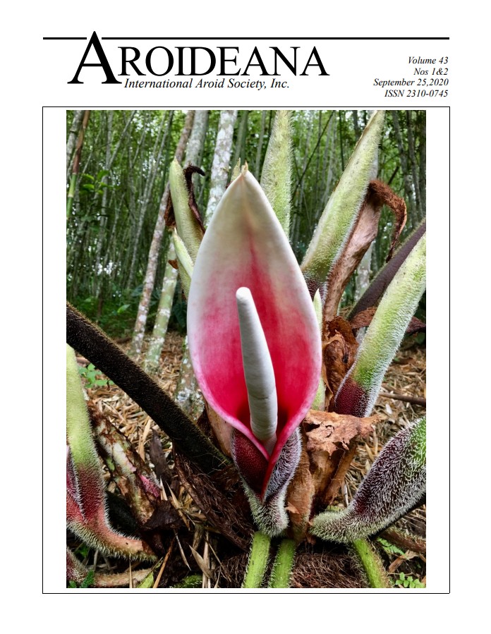
anatomy of flowers and resiniferous canals of Philodendron ornatum (Araceae)
Denis Barabe,
Kshitij Trivedi, & Christian Lacroix
ABSTRACT
This study
documents different stages of development of inflorescences and floral types
(staminate, pistillate, sterile male, and atypical bisexual flowers) in Philodendron
ornatum from an anatomical perspective, to track
the development and structure of resiniferous canals, raphide
idioblasts, and laticifers. Resiniferous canals and
laticifers develop during early stages of initiation of floral primordia
predominantly in the male and sterile male floral zones. As floral organs like
stamens and staminodes are initiated, resiniferous canals become more apparent
and line the area immediately below those organs. There are comparatively fewer
resiniferous canals in the intermediate zone of the spadix. The vascular
bundles are surrounded by laticifers throughout the axis of the inflorescence and
raphide idioblasts are
scattered throughout the aerenchymatous ground tissue.
Mature resiniferous canals consist of one or two cell layers surrounding a
central cavity. The internal layer corresponds to the epithelial cells that
release resin. This type of canal corresponds to the more common type described
in other species. Resiniferous canals were also observed with much less frequency
on the outer periphery of the ground tissue just below the gynoecia of some
female flowers. We conclude that there is a need to broaden this type of study to
include a survey of more species to determine if there is a strong correlation between
the anatomy of resiniferous canals and the phylogeny of species of Philodendron
and Thaumatophyllum.
KEYWORDS
Resiniferous
canals, inflorescence, anatomy, floral development, Philodendron ornatum, laticifers, raphides.